Last updated on April 4th, 2025 at 02:33 pm
Neurons constitute the basic part of our nervous system. They are a key element in the manner in which our body captures, analyzes, and gives response to any information. Knowing the anatomy of a neuron enables one to understand how our brain and nervous system function.
What Is a Neuron?
A neuron is also referred to as a nerve cell, and it serves to be the structural and functional unit of the nervous system. Its role includes receiving, processing, and sending chemical and electrical signals across the body. The basic unit of communication in the nervous system is synapse which allow for smooth action in movement, sensation, thinking, and other bodily functions working together seamlessly.
What sets neuron apart is its inability to form anew as easily as other body cells. Therefore, it is imperative to case and earn our nervous system health.
Neuron Structure and Function
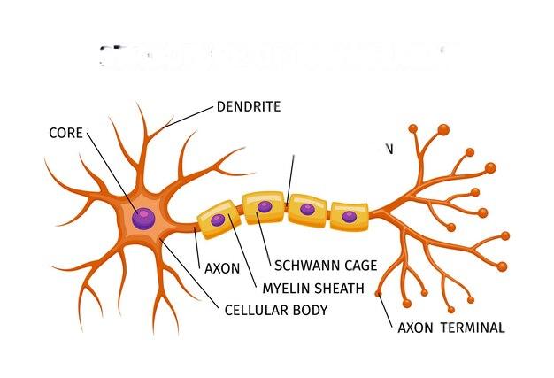
A neuron consists of several specialized structures that help it perform its function. Let’s explore the parts of a neuron and their roles:
1. Dendrites
- Short, branching extensions of the neuron
- Receive incoming signals from other neurons
- Transmit signals toward the cell body
2. Cell Body (Soma)
- The central part of the neuron that contains the nucleus
- Processes incoming information from dendrites
- Responsible for cellular metabolism and maintenance
3. Axon
- A long, tube-like extension that carries electrical signals away from the cell body
- Can be covered in a myelin sheath, which speeds up signal transmission
4. Myelin Sheath
- A fatty layer that insulates the axon
- Speeds up nerve impulses, ensuring quick and efficient communication
5. Axon Terminals
- The branching ends of the axon that connect with other neurons, muscles, or glands
- Release neurotransmitters, chemical messengers that cross the signals between neurons.
A neuron diagram helps imagine these structures, making it easy to understand how they function together.
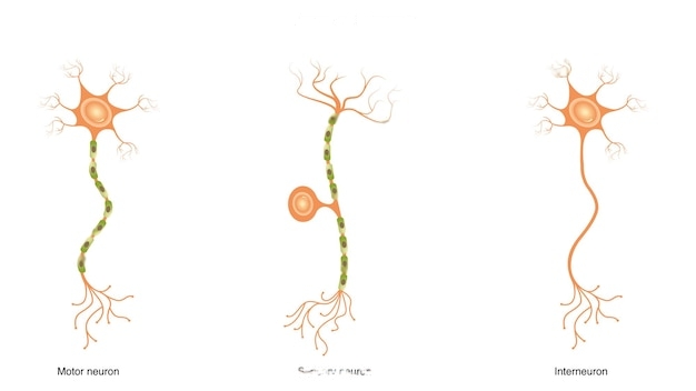
Types of neurons
Neurons come in different types, each is special for a unique function. There are three main types of neurons:
1. Sensory Neurons
- Found in peripheral nervous system.
- Find out external stimuli (eg : touch, temperature, pain) and send signals to the central nervous system (CNS)
- Example: Nerve endings in the skin which is a feeling of heat.
2. Motor Neurons
- Control muscle movements by transmitting signals from the CNS to muscles or glands
- Essential for voluntary and involuntary movements
3. Interneurons
- Found in the CNS (brain and spinal cord)
- Act as connectors, relaying signals between sensory and motor neurons
A nerve cell diagram often distinguishes these types of neurons by showing their distinct structures and pathways.
Nervous System Parts: How Neurons Fit In
The nervous system is a huge network of neurons working together. It is divided into two main parts:
1. Central nervous system (CNS)
- Consists of Brain and spinal cord
- Processes and explains sensory information
- Sends signals to start action
2. Peripheral nervous system (PNS)
- These consists of nerves outside the brain and spinal cord
- Further divided into somatic nervous system (voluntary movements) and autonomic nervous system (involuntary functions like heartbeat and digestion)
The peripheral nervous system plays an important role in connecting the body to CNS, allowing movement, sensation and reactions to stimuli.
Neuron Picture: A Visual Representation
A neuron picture or diagram is an essential teaching tool that simplifies the understanding of the neuron anatomy. This clearly laces parts of a neuron, showing how to travel from signal dendrite to exon terminals. Looking at a neuron diagram makes it easy to understand how neurons interact within the nervous system.
Neuron Function: How Neurons Work in the Nervous System
Neurons communicate using electrical and chemical signals, causing rapid transmission of information across the body. His primary function is:
- Get information from sensory receptors or other neurons.
- Process and integrate information within the central nervous system.
- Send signals to muscles, glands, or other neurons to generate reaction.
This complex process ensures that we can think efficiently, move and react to our environment.
Electrical Transmission: The Action Potential
Neurons generate and transmit signals through electrical impulses, which are called action potential. This process takes place in several stages:
1. Resting Potential: The Neuron at Rest
- A neuron at rest has a slightly more negative charge inside than outside.
- This charge is maintained by ion pumps that regulate sodium (NA⁺) and potassium (K⁺) levels.
2. Stimulus & Threshold: The Triggering Event
- When a neuron receives a strong enough signs, it reaches a threshold level.
- This causes an opening on sodium channels, allowing Na⁺ to rush in the neuron.
3. Depolarization: The Action Potential Begins
- The influx of Na⁺ changes the charge from negative to positive inside the neuron.
- This electrical impulse takes the axon down like a wave.
4. Repolarization: Resetting the Neuron
- Potassium (K⁺) channels opens, allowing K⁺ to leave, the negative charge is restored.
- Neuron returns to its rest position, which is ready for the next signal.
5. Signal Transmission to the Next Neuron
- Once the action potentials reach the axon terminals, it triggers the release of the neurotransmitters into the synapse.
- These chemicals take the message to the next neuron or muscle cell.
The entire process is only in the millisecond, which gives rapid reactions to stimuli.
Chemical Transmission: The Role of Neurotransmitters
While neurons use electrical impulses to carry signals with their length, they use chemical messengers to pass the signal to the next neuron or target cell. It occurs on special junctions called synapses.
Steps in Chemical Transmission
1. Electric signal reaches the Axon terminal.
2. It triggers the release of neurotransmitters (chemical messengers).
3. Neurotransmitters cross the synaptic gap and bind receptors on the next neuron.
4. The obtained neuron generates a new electrical signal if stimulation is strong enough.
Types of Neurotransmitters & Their Functions
| Neurotransmitter | Function | Example of Effect |
| Acetylcholine (ACh) | Muscle movement, learning | Controls muscle contractions |
| Dopamine | Pleasure, reward, motivation | Affects mood & voluntary movement |
| Serotonin | Mood regulation, sleep | Linked to depression & happiness |
| GABA (Gamma-Aminobutyric Acid) | Inhibitory neurotransmitter | Reduces brain activity, prevents overstimulation |
| Glutamate | Excitatory neurotransmitter | Enhances learning & memory |
FAQs Neuron Anatomy 101: Diagrams & Simple Explanations
1. What does a neuron look like?
Answer: A neuron has a star -shaped cell body (Soma), with a branch -like dendrites that extend outwards. It also contains a long axon that ends the signal in axon terminals which connects to other neurons. Some axons are covered with a myelin sheath, which speeds up signal transmission. A neuron diagram represents these parts visually.
2. How many neurons are there in the human body?
Answer: The human body consists of about 86 billion neurons, which are mostly concentrated in the brain and spinal cord. There are also millions of neurons in the peripheral nervous system that connect the brain to muscles and organs. These neurons create giant networks enabling complex ideas, movements and sensations. The exact number differs slightly between individuals.
3. How do neurons communicate with each other?
Answer: Neurons communicate through electrical impulses (action potential) and chemical signals (neurotransmitters) on the synapses. Electrical signals move the axon down and trigger the release of the neurotransmitter. These chemicals cross the synaptic gap to stimulate the next neuron.
4. What are different types of neurons?
Three main types of neurons sensory neurons (carrying signs from sensory organs to brain), motor neurons (sending commands from brain to muscles), and interneurons (connect neurons within the brain and spinal cord). Each nervous system plays a unique role in function.
5. Why is myelin sheath important?
Answer: Myelin sheath is a fatty layer that insulations the axon, allowing nerve impulses to travel faster. This helps improve communication between neurons and prevents signal loss. Damage to myelin sheath, as seen in diseases such as multiple sclerosis, slows down nerve signaling.